Back Muscles Anatomy Labeled - Anatomy Posters Full Circle School Of Massage Therapyfull Circle School Of Massage Therapy / This chapter is divided into three main sections:
Back Muscles Anatomy Labeled - Anatomy Posters Full Circle School Of Massage Therapyfull Circle School Of Massage Therapy / This chapter is divided into three main sections:. This chapter is divided into three main sections: Anatomical diagram showing a back view of muscles in the human body. Within this group of back muscles you will find the latissimus dorsi, the trapezius, levator scapulae and the rhomboids. Learn about anatomy muscle labeling with free interactive flashcards. The superficial back muscles are the muscles found just under the skin.
These muscles are able to move the upper limb as they originate at the vertebral column and insert onto. Learn about anatomy muscle labeling with free interactive flashcards. The extrinsic muscles include the trapezius, latissimus dorsi, rhomboid major and minor, levator scapulae and the serratus posterior superior and. The muscles of the back are divided into two groups, the extrinsic and the intrinsic muscles, which the intrinsic back muscles are sequestered in an osseofibrous canal, formed by the thoracolumbar fascia, the vertebral arches, and the spinous and transverse processes of associated vertebrae. The back muscles can be three types.
Muscle Diagram Of The Back Posterior Front Anterior from www.alpha-athlete.com Muscle anatomy quiz for anatomy and physiology! This site was designed for students of anatomy and physiology. These muscles give height and breadth to back. They are divided into three groups, as shown below. Included are several layered views of the back muscles, the doral muscles, subclavius muscles, rhomboideus major and minor muscles, deltoid muscles and many more. Topographically, the muscles in this group are classed along with the lateral torso wall and upper extremity, which is due to their location as well as their genetic development based on their embryological origin. Their main function is contractibility. The muscular system is responsible for movement in collaboration with the nervous system to form impulses for motion.
Muscles, connected to bones or internal organs and blood vessels, are in charge for movement.
Muscles, connected to bones or internal organs and blood vessels, are in charge for movement. It contains textbook resources, such as chapter review guides, homework sets, tutorials chapter 8: The muscular system is responsible for movement in collaboration with the nervous system to form impulses for motion. The superficial back muscles are the muscles found just under the skin. Click on the labels below to find out more about your muscles. Muscle basics and cellular components, naming of the muscles, and cat. Tutorials on the anatomy and actions of the back muscles, using interactive animations, diagrams, and illustrations. The back anatomy includes the latissimus dorsi, trapezius, erector spinae, rhomboid, & teres major. The muscles of the shoulder and back chart shows how the many layers of verified purchase. This chapter is divided into three main sections: Muscles also contribute to internal functions of the human body which include motion in the intestines and circulatory system. While working on neck muscles. This article covers the anatomy of the deep muscles of the back, including their function, blood supply, innervation, origin and insertion.
This is a table of skeletal muscles of the human anatomy. Anatomical diagram showing a back view of muscles in the human body. This chapter is divided into three main sections: Muscle basics and cellular components, naming of the muscles, and cat. They are located deep to the extrinsic muscles, being separated from them by the thoracolumbar fascia.
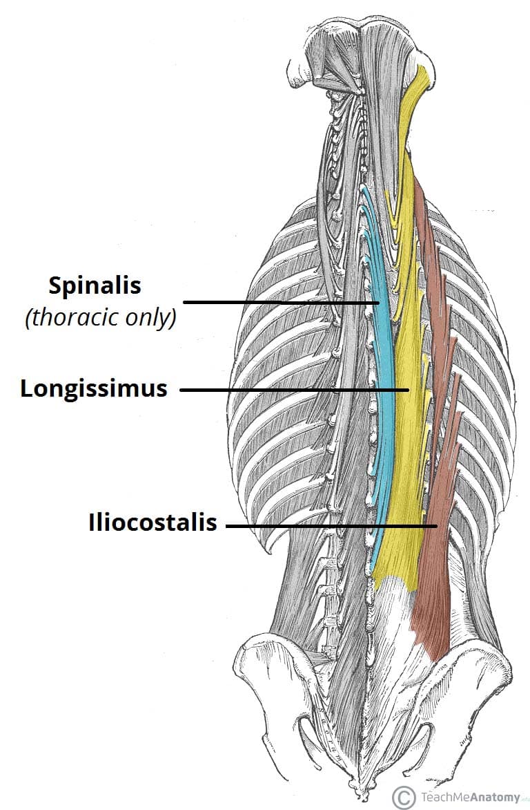
Muscles Of The Back Teachmeanatomy from teachmeanatomy.info They are located deep to the extrinsic muscles, being separated from them by the thoracolumbar fascia. Their main function is contractibility. This site was designed for students of anatomy and physiology. Included are several layered views of the back muscles, the doral muscles, subclavius muscles, rhomboideus major and minor muscles, deltoid muscles and many more. 12 photos of the muscle anatomy labeled. Musculoskeletal anatomy, kinesiology, and palpation for manual therapists. Muscles, connected to bones or internal organs and blood vessels, are in charge for movement. While working on neck muscles.
These muscles are able to move the upper limb as they originate at the vertebral column and insert onto.
Within this group of back muscles you will find the latissimus dorsi, the trapezius, levator scapulae and the rhomboids. Choose from 500 different sets of flashcards about anatomy muscle labeling on quizlet. When you are taking anatomy and physiology you will be required to identify major muscles in the human body. By jholmesrn419 , may 2007. Intermediate back muscles and c. Learn about anatomy muscle labeling with free interactive flashcards. The muscles of the back are divided into two groups, the extrinsic and the intrinsic muscles, which the intrinsic back muscles are sequestered in an osseofibrous canal, formed by the thoracolumbar fascia, the vertebral arches, and the spinous and transverse processes of associated vertebrae. These muscles are able to move the upper limb as they originate at the vertebral column and insert onto. It contains textbook resources, such as chapter review guides, homework sets, tutorials chapter 8: Human muscle system, the muscles of the human body that work the skeletal system, that are under voluntary control, and that are concerned with the following sections provide a basic framework for the understanding of gross human muscular anatomy, with descriptions of the large muscle groups. Microscopic anatomy of skeletal muscle. Learn about these muscles, their locations there are several individual muscles within the back anatomy, and it's important to take a quick look at all of them to see how you can target them. Clearly pictured and labeled muscles of the back and neck.
12 photos of the muscle anatomy labeled. Exercise of this organ system is critical to prevent. The deep back muscles lie immediately adjacent to the vertebral column and ribs. The superficial back muscles are the muscles found just under the skin. This chapter is divided into three main sections:
Male Back Anatomy Anatomy Drawing Diagram from images-na.ssl-images-amazon.com The muscles of the back are separated into extrinsic and intrinsic components, which are based on their function in movement and embryological origin. Microscopic anatomy of skeletal muscle. This site was designed for students of anatomy and physiology. 12 photos of the muscle anatomy labeled. By jholmesrn419 , may 2007. The muscles of the shoulder and back chart shows how the many layers of verified purchase. When you are taking anatomy and physiology you will be required to identify major muscles in the human body. Their main function is contractibility.
Click on the labels below to find out more about your muscles.
They are divided into three groups, as shown below. Learn about anatomy muscle labeling with free interactive flashcards. By jholmesrn419 , may 2007. This chapter is divided into three main sections: These muscles give height and breadth to back. The extrinsic muscles include the trapezius, latissimus dorsi, rhomboid major and minor, levator scapulae and the serratus posterior superior and. 12 photos of the muscle anatomy labeled. Included are several layered views of the back muscles, the doral muscles, subclavius muscles, rhomboideus major and minor muscles, deltoid muscles and many more. Memorize all the muscle facts with the help of muscle cheat sheets. Microscopic anatomy of skeletal muscle. Muscles, connected to bones or internal organs and blood vessels, are in charge for movement. The muscles of the back are separated into extrinsic and intrinsic components, which are based on their function in movement and embryological origin. Learn about these muscles, their locations there are several individual muscles within the back anatomy, and it's important to take a quick look at all of them to see how you can target them.
The muscles of the back are separated into extrinsic and intrinsic components, which are based on their function in movement and embryological origin back muscles anatomy. Within this group of back muscles you will find the latissimus dorsi, the trapezius, levator scapulae and the rhomboids.


Posting Komentar
0 Komentar