Anterior Shoulder Tendon Anatomy : Posterior muscles and ligaments of the shoulder girdle ... : Tendons are fibrous cords attached to muscles and bone.
Anterior Shoulder Tendon Anatomy : Posterior muscles and ligaments of the shoulder girdle ... : Tendons are fibrous cords attached to muscles and bone.. Specifically, the four rotator cuff muscles include the following This webpage presents the anatomical structures found on shoulder mri. Glenohumeral joint glenohumeral joint the glenohumeral joint is a multiaxial synovial ball and socket joint and involves articulation between the glenoid fossa of the. Start studying anterior shoulder anatomy. 3 405 337 просмотров 3,4 млн просмотров.
Robin smithuis and henk jan van der woude. Normal anatomy, variants and checklist. The synovial part of the tendon sheath consists of a visceral and parietal layer separated by synovial fluid. Glenohumeral joint glenohumeral joint the glenohumeral joint is a multiaxial synovial ball and socket joint and involves articulation between the glenoid fossa of the. Flexes and medially rotates arm;
MRI of Shoulder anatomy from image.slidesharecdn.com Corey chakarun from shin imaging in california. Start studying anterior shoulder anatomy. 3 405 337 просмотров 3,4 млн просмотров. • pain and/or pop at anterior shoulder but usually not painful after initial event. Latarjet procedure performed more commonly than bristow. In addition, the trapezius, serratus anterior, rhomboids, and levator scapulae insert on the scapula and are responsible for the muscles and tendons of the rotator cuff form a cover around the anterior, superior, and posterior humeral head. There are also fibrous bands, known as retinacula, which make a tunnel around the. Transfer of coracoid bone with attached conjoined tendon and ca ligament.
3 405 337 просмотров 3,4 млн просмотров.
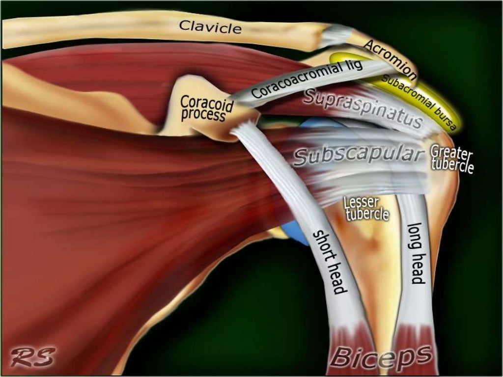
Shoulder anatomy is an elegant piece of machinery having the greatest range of motion of any joint in the body. The pectoralis minor muscle is a small. Scapula and related structures — the scapula is a relatively large, flat bone located on the posterior thorax (figure 1 and the anterior and posterior portions of the supraspinatus muscle give rise to distinct portions of the supraspinatus tendon. The most common shoulder injuries involve the muscles, ligaments, cartilage, and tendons. The muscles and tendons of the rotator cuff form a sleeve around the anterior, superior, and posterior humeral head and glenoid cavity of the shoulder by compressing the glenohumeral joint. The shoulder muscles are associated with movements of the upper limb. 1 enumerate the layers of anterior abdominal wall. • pain and/or pop at anterior shoulder but usually not painful after initial event. Corey chakarun from shin imaging in california. The human shoulder is made up of three bones: Biceps brachii origin (proximal attachment). 3 405 337 просмотров 3,4 млн просмотров. Anterior graphic of the shoulder.
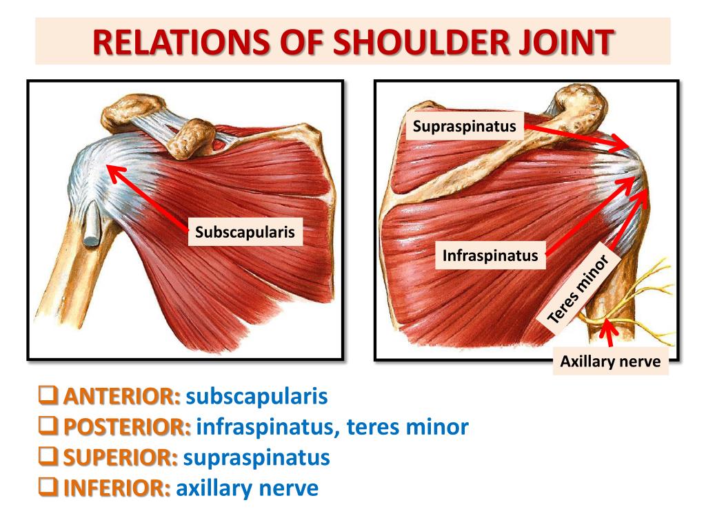
Most common finding is 'military patch' (deltoid) anesthesia. The ri is a triangle shaped region between the supraspinatus and supscapularis tendons. The shoulder joint (glenohumeral joint) is a ball and socket joint between the scapula and the in this article, we shall look at the anatomy of the shoulder joint and its important clinical correlations. Upper limb trauma programme injuries. Anterior graphic of the shoulder.
PPT - ANATOMY OF THE SHOULDER REGION PowerPoint ... from image1.slideserve.com The tendon of the subscapularis muscle attaches both to the lesser tubercle aswell as to the greater tubercle giving. Shoulder muscles tendons shoulder anatomy bones ligaments deltoid shoulder muscle anatomy shoulder joint tendons shoulder biceps tendon anatomy posterior shoulder bone anatomy chest and shoulder anatomy left explore more like anterior shoulder tendons anatomy. Infraspinatus and teres minor tendon. A fibrous layer, made of tight collagenous tissue, and a synovial layer. • pain and/or pop at anterior shoulder but usually not painful after initial event. Tendon sheaths consist of two layers: Majority of anterior shoulder dislocations are due to trauma. Corey chakarun from shin imaging in california.
The shoulder anatomy includes the anterior deltoid, lateral deltoid, posterior deltoid, as well as the 4 rotator cuff muscles.
In the shoulder, articular cartilage covers the end of the humerus and socket area of the glenoid on the scapula. Subscapularis tendon (open arrow) and anterior labrum (arrowhead) are also shown on this section. The synovial part of the tendon sheath consists of a visceral and parietal layer separated by synovial fluid. Important to rule out axillary nerve injury. Anterior static shoulder stability is provided by. 3 405 337 просмотров 3,4 млн просмотров. Simple, easy notes for quick revision of important questions. 2 name the planes used for dividing abdominal cavity into regions. Irreducible anterior dislocation of the shoulder due to interposition of the long head of bíceps tendón and avulsed part of the labrum, treated arthroscopically; The tendons that control movement in your hands, wrists and fingers run through your forearm. Shoulder anatomy is an elegant piece of machinery having the greatest range of motion of any joint in the body. Anterior graphic of the shoulder. Glenohumeral joint glenohumeral joint the glenohumeral joint is a multiaxial synovial ball and socket joint and involves articulation between the glenoid fossa of the.
Majority of anterior shoulder dislocations are due to trauma. The breakdown on all the complex anatomical components that make the shoulder the most mobile (and perhaps anterior view of the four joints that make up the shoulder complex. Irreducible anterior dislocation of the shoulder due to interposition of the long head of bíceps tendón and avulsed part of the labrum, treated arthroscopically; Latarjet procedure performed more commonly than bristow. Anterior part of the deltoid:
Illustration of the relevant measured neurovascular ... from www.researchgate.net The tendons that control movement in your hands, wrists and fingers run through your forearm. The pectoralis minor muscle is a small. A fibrous layer, made of tight collagenous tissue, and a synovial layer. Start studying anterior shoulder anatomy. Shoulder anatomy for ultrasound evaluation. • review pertinent anatomy and pathology associated with common causes of shoulder pain. Robin smithuis and henk jan van der woude. The etiology is most of the time traumatic and related either to sport or accidents.
Dynamic anterior shoulder stabilization with the long head of the biceps tendon:
One of the most visible and important tendons in this area is the biceps tendon which attaches the biceps muscle to. Posterior part of the deltoid: In the shoulder, articular cartilage covers the end of the humerus and socket area of the glenoid on the scapula. • review pertinent anatomy and pathology associated with common causes of shoulder pain. Subscapularis tendon (open arrow) and anterior labrum (arrowhead) are also shown on this section. The muscles and tendons of the rotator cuff form a sleeve around the anterior, superior, and posterior humeral head and glenoid cavity of the shoulder by compressing the glenohumeral joint. Start studying anterior shoulder anatomy. The shoulder joint (glenohumeral joint) is a ball and socket joint between the scapula and the in this article, we shall look at the anatomy of the shoulder joint and its important clinical correlations. The tendons that control movement in your hands, wrists and fingers run through your forearm. Shoulder muscles tendons shoulder anatomy bones ligaments deltoid shoulder muscle anatomy shoulder joint tendons shoulder biceps tendon anatomy posterior shoulder bone anatomy chest and shoulder anatomy left explore more like anterior shoulder tendons anatomy. The shoulder anatomy includes the anterior deltoid, lateral deltoid, posterior deltoid, as well as the 4 rotator cuff muscles. 2 name the planes used for dividing abdominal cavity into regions. Scapula and related structures — the scapula is a relatively large, flat bone located on the posterior thorax (figure 1 and the anterior and posterior portions of the supraspinatus muscle give rise to distinct portions of the supraspinatus tendon.
In the shoulder, articular cartilage covers the end of the humerus and socket area of the glenoid on the scapula shoulder tendon anatomy. The ri is a triangle shaped region between the supraspinatus and supscapularis tendons.


Posting Komentar
0 Komentar